

Overview
Read this article from a comprehensive knowledge base, updated and supplemented with articles reviewed by scientific committees.
Read the articleAUTHOR
-
Gérard ROBLIN : Doctor of Science - Director of Research at the Centre national de la recherche scientifique (CNRS) (ER)
INTRODUCTION
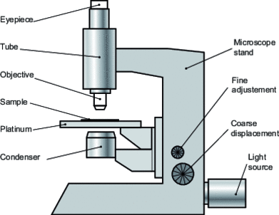
The optical microscope is essentially made up of two optical components: the objective and the eyepiece (figure ). This assembly, attached to a tube, is pointed at the object or preparation placed on a table or stage equipped with devices for positioning the object in its plane, generally by two translational movements and/or one rotational movement. Focusing is ensured by two translation stages parallel to the optical axis: fast and slow movements (highly sophisticated instruments used in research laboratories may have an ultra-slow movement, while some teaching microscopes may only have a single movement with an amplitude and speed intermediate to the usual values). These various elements are linked together, and their stability ensured, by a mechanical mount, the stand, for which we can often distinguish a foot and a stem. Illumination of the object is provided by a condenser and source assembly, this one is most often also attached to the stand nowadays (and housed in its base.) This illumination can take different forms, depending on the type of observation required for the object.
But an instrument would not be complete without a receiver to collect the information, i.e. to capture the image it provides. As the optical microscope is capable of providing images using visible or near-infrared and ultraviolet light (for wavelengths between 0.22 and 1.7 µm), the observer's eye cannot always be the direct receptor, and intermediates such as photographic emulsion, fluorescent screen or electronic image converter must be used, all of which justify the possible presence of several "image outputs", enabling the image to be preserved, distributed and collectively observed.

Exclusive to subscribers. 97% yet to be discovered!
Already subscribed? Log in!
Light microscopy
 Geometrical optics of the microscope
Bibliography
General books and articles
Exclusive to subscribers. 97% yet to be discovered!
Already subscribed? Log in!
Article included in this offer
"Mechanical and dimensional measurements"
(
126 articles
)
Updated and enriched with articles validated by our scientific committees
A set of exclusive tools to complement the resources
